HOW IT WORKS
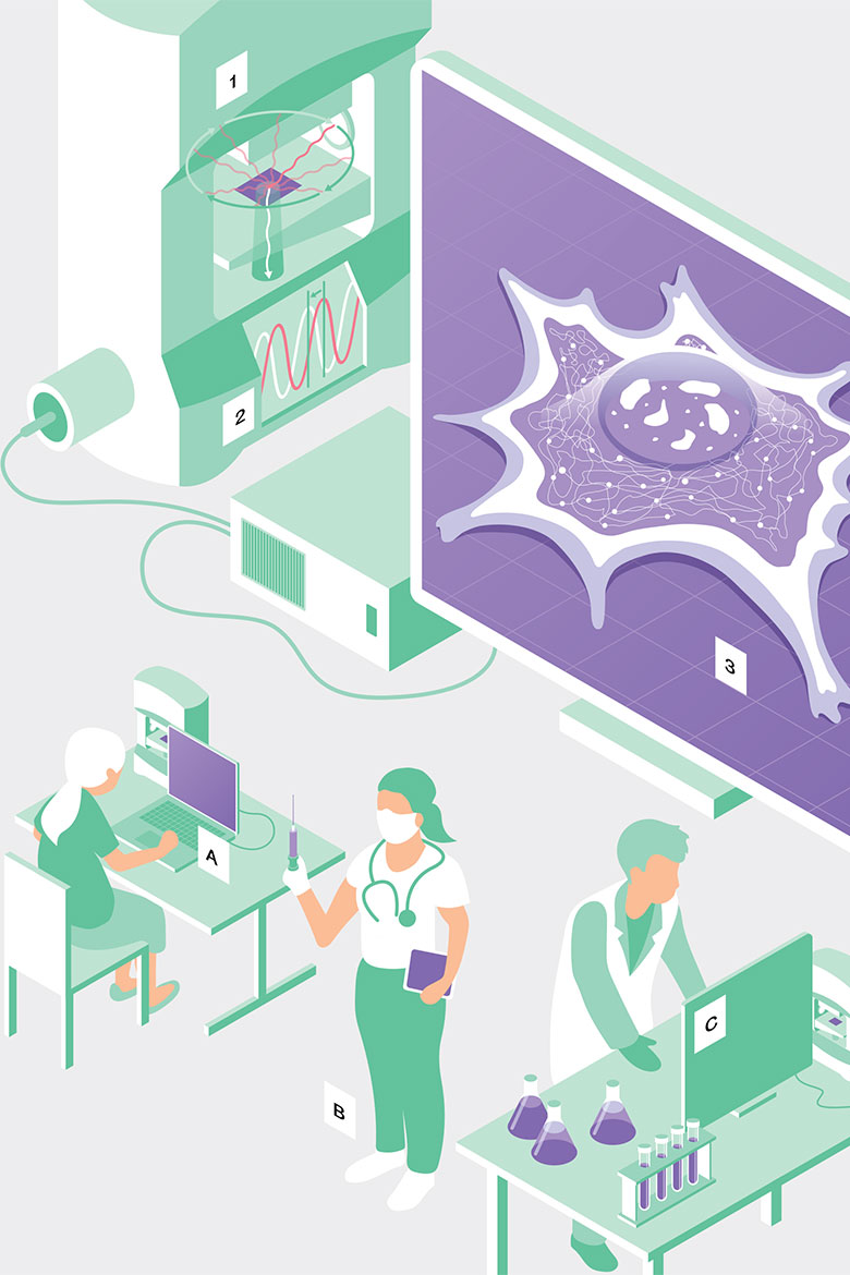
A living cell becomes a hologram
Microscopes have always been a determining factor for progress in the field of biology. An EPFL spin-off now uses a laser to look deep into the interior of cells and create a 3D picture of them.
Traditional light microscopes soon reach their limits. You can only see large-scale structures inside cells, because their radiation kills the living material, and the image is only two-dimensional. Nanolive, an EPFL spin-off, has now developed an innovative microscope that can take things a step further.
To find out how it works, hover your mouse over the numbers and letters, or tap on them with your finger.

Suitable for varied purposes
(A) In schools: The cells don’t have to be prepared especially, and the device is simple to use.
Suitable for varied purposes
(B) For diagnostic purposes: The microscopes don’t use harmful radiation or colouring chemicals, so they are suitable for investigating the health of embryos to be used in artificial fertilisation.
Suitable for varied purposes
(C) In research: Cells can be observed over a longer period of time without being destroyed. They can be watched as they divide, for example, or while they communicate with neighbouring cells and react to drugs.
The device shoots a weak laser at the living cells. A rotating mirror ensures that the light falls on the sample from all sides.
Most cells are transparent and hardly offer any contrast, so the microscope has a trick up its sleeve: the oscillations of the laser light are hindered in different ways by different components of the cell (this is called ‘phase-shifting’). Information can be gained by superimposing the light waves before and after they emerge out of the sample.
Software is then used to calculate a hologram from the overlayered laser. It shows delicate structures within a cell, in three dimensions. The cells are still alive in the microscope, which means their movements can be captured.
Illustration: ikonaut