Weaker magnets, better pictures
Tech downgrades can make MRI applications cheaper and more flexible

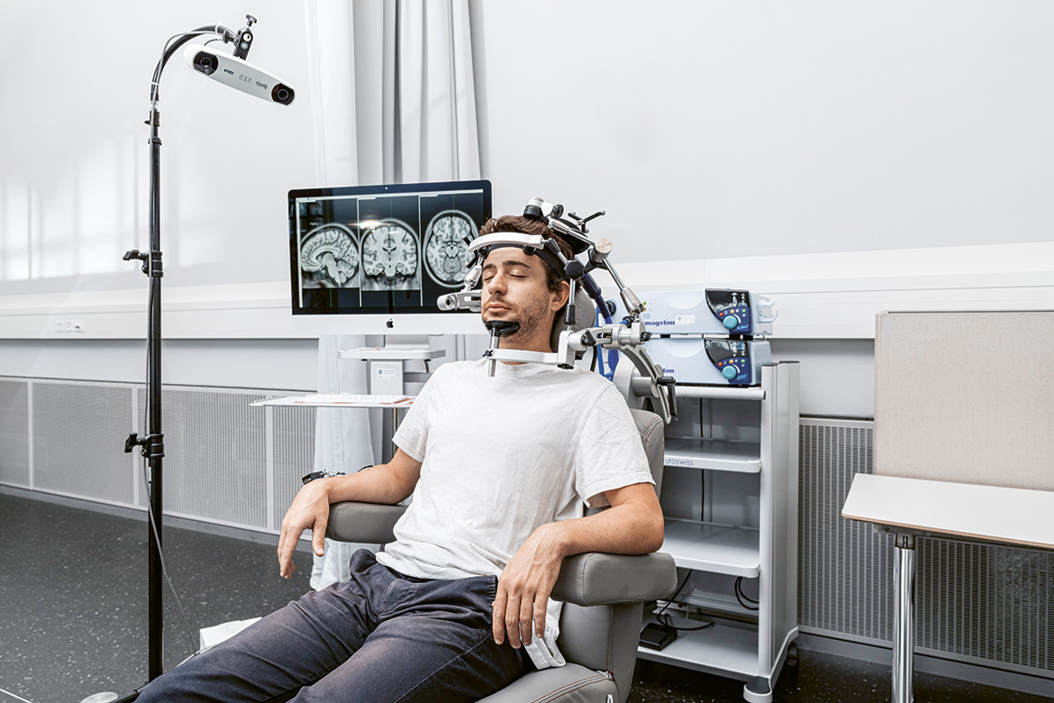
Downsizing in medicine: Researchers at the University of Basel are working with prototypes of small MRI machines. | Image: University of Basel, photographer: Donata Ettlin
Whether someone is suffering from a stroke, cancer or a sports injury, magnetic resonance imaging (MRI) is becoming ever more vital in helping doctors to make a more precise diagnosis. The magnets in these machines have been getter bigger and bigger in recent years. Today, some hospitals even have MRI machines with a field strength of seven tesla – that’s more than 100,000 times stronger than the Earth’s magnetic field. The stronger the magnet, the more detailed the images become, without any need to increase the time spent on an examination.
But this progress comes at a cost. Modern MRI machines cost millions, and weigh several tons. They need specially shielded rooms and an expensive cooling system using liquid helium – a resource that is becoming ever scarcer. What’s more, people with implants cannot have an MRI involving 1.5 tesla or more, because it might heat up the electrodes of their heart pacemakers or hip replacements, and lead to image distortions.
Small machines offer greater mobility
This is why physics professor Najat Salameh and her colleague Mathieu Sarracanie have been taking things in the opposite direction. At the Department of Biological Engineering of the University of Basel they have been developing machines that aim to function with far weaker magnetic fields. Their goal is to make small MRI machines that are flexible enough to be adapted to different needs. This would make it possible to employ them in regions that are difficult to access, in countries without any regular electricity supply, and on patients with implants. “We don’t see ourselves in competition with the high-performance MRI machines, but as a valuable complement to them”, says Salameh.
Salameh and Sarracanie have already built several prototypes with magnetic fields ranging from 0.01 to 0.1 tesla, which removes the necessity for elaborate shielding and helium cooling. At present, they are using electromagnets driven by the mains supply, but one day they could function using batteries too. And they are small enough to fit into an ambulance or an off-road vehicle.
Profiting from technological progress
However, the big question is whether or not these weaker magnetic fields can also produce high-quality images. Najat Salameh is convinced they can: “Twenty years ago, work on MRIs with weak magnets was practically abandoned in favour of those with strong magnets”. But immense progress has been made since then, both in matters of technology and in data processing.
Sebastian Kozerke is a professor of biomedical imaging at ETH Zurich and the University of Zurich. He, too, thinks it is fundamentally right and proper to resume research into weaker field strengths. MRI machines are today largely confined to highly developed countries because of their high costs and the infrastructure necessary. “We have to look for ways to compromise and to reduce the field strength enough to lower costs, while ensuring that the image quality remains sufficient”, he says. But he is sceptical about whether MRIs using a magnetic field of below 0.1 tesla – such as are currently in development – will ever deliver the required results without recourse to expensive signal amplification.
But Salameh and Sarracanie are taking an approach that seems promising. Their image resolution using weak magnetic fields is not yet comparable with other, modern MRI machines, but it offers a higher degree of contrast. This makes it easier to differentiate between various types of tissue – whether healthy or diseased. What’s more, they are planning to optimise further components one at a time, such as signal detection and data processing. If this is successful, many more people might in future be able to benefit from an MRI examination than is presently the case.
A glimpse into the interior